Copyright (c) 1999 by Today Enterprises.
Article published in Microscopy Today 99-6: 22-24 (1999).
http://microscopy-today.com/PDFFiles/MT-1999-06-small.pdf
Reproduced with permission.
______________________________________
A thin, flat section of animal or plant tissue will usually adhere to a clean glass surface and stay in place through the common preparative procedures of staining, washing, dehydration and clearing. The adhesion may be due largely to the close contact between two flat surfaces. This allows non-ionic forces (such as van der Waals forces and hydrogen bonds), which are very weak between individual atoms, to exist in numbers great enough to add up to a strong attraction between slide and section. Ionic attractions, which operate over longer distances, may also exist between the silicate of the glass (negatively charged ions, especially in an alkaline medium) and basic groups of proteins in the tissue (positive ions, especially in an acidic medium). Unfortunately, not all sections are perfectly flat and not all slides are perfectly clean. Even when these conditions are met, there are some staining techniques that are harsh enough to remove the flattest section from the cleanest slide.
There are two ways to prevent or reduce the loss of sections subjected to harsh treatments: (a) by using an adhesive, and (b) by inclusion in a permeable protective film.
Adhesives.
The most popular way to try to keep sections on slides is to coat the glass with an adhesive. Many kinds are available and some are now briefly reviewed. The efficiency of an adhesive depends on the nature of the treatment that might cause detachment of the sections.
Adhesion of oppositely charged surfaces.
When immersed in an aqueous medium in the pH range 5 to 7, most animal tissues carry a net negative charge, owing to a small excess of acidic over basic amino acids in the structural proteins. Plant tissues are composed largely of cellulose, which does not itself form ions but is impregnated with a variety of weak acids, and these too confer an overall negative charge. Sections of almost any tissue can therefore be expected to adhere well to a glass surface that has been treated in such a way as to make it positively charged. The modification is usually accomplished either by coating the slide with a basic polymer or by a chemical reaction that leaves amino groups linked by covalent bonds to the silicon atoms of the glass.
A frequently used basic polymer is polylysine, in which every amino acid unit has an amino side chain that is quite strongly basic. Its pKa is 10.53. This is the pH at which half the amino groups are protonated. The side-chain of lysine has an unusually high pKa for an amino group and consequently polylysine is positively charged even in moderately alkaline media. Polylysine is applied to slides as an aqueous solution, which is then allowed to dry by evaporation. Either D- or L-polylysine or the somewhat less expensive racemic DL- mixture may be used because the stereochemistry does not affect the ionization of the side chain. The guanidine side-chain of arginine is a strong base (pKa=12.5). Polyarginine should therefore retain its positive charge even in the most strongly alkaline conditions, but it is even more expensive than polylysine and does not seem to have been used as a section adhesive.
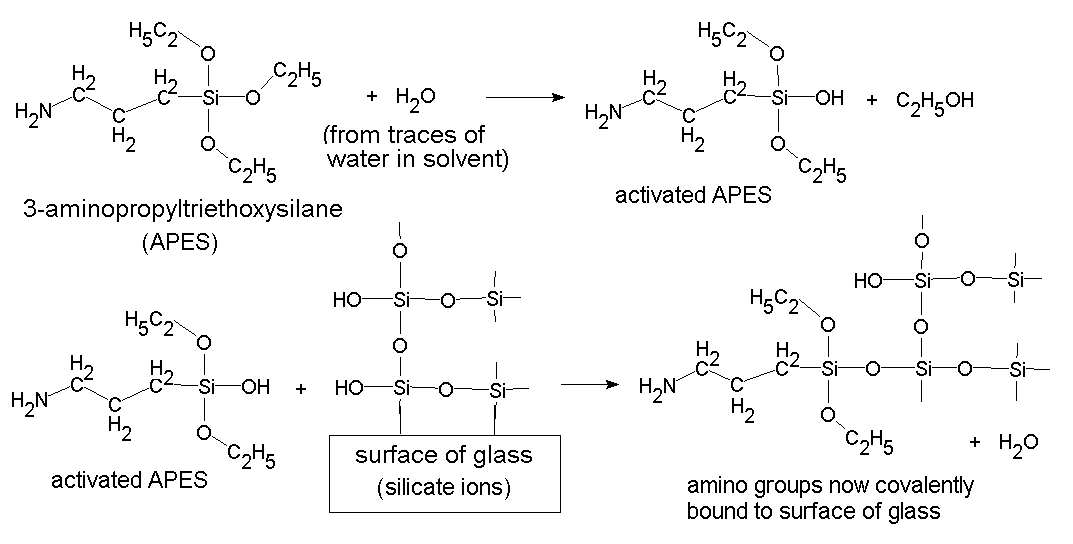
The other way to make a positively charged glass surface is to treat it with a solution of 3-aminopropyltriethoxysilane (APES) in the presence of catalytic traces of water. The 0.1% or so of water ordinarily present in acetone is enough.
The positive charge of APES-treated (silanized) slides is like that imparted by polylysine, but covalent bonding to the glass ensures that it cannot be washed away.
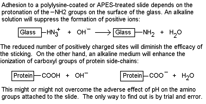
The carboxyl groups of proteins (and of some macromolecular carbohydrates) vary in their responses to pH. All are weak acids: many remain ionized at pH 2.5 but all are uncharged at pH 1.
Some tissues (cartilage matrix is a notable example) contain an abundance of sulphate-ester groups. These are strong acids, ionized at any pH. Tissues of this kind are therefore attracted to protonated amino groups however low the pH, but will not be attracted by a negatively charged surface such as glass with its silicate ions or an adhesive coating that contains an excess of carboxyl groups.
BOX 1 contains instructions for a simple procedure that attaches amino groups to the surfaces of glass slides.
----------------------------------------
BOX 1. Making silanized slides.
What you need:
1. Slides (must be very clean, and dry)
2. Staining racks to hold the slides (glass or metal)
3. Staining tanks (one just large enough to hold the staining rack, and one of larger size)
4. Water (distilled or deionized): Fill the larger staining tank
5. Silanizing reagent. Add 4 ml of 3-amino-propyltriethoxysilane to 200 ml of acetone in the small staining tank.
This solution must be used on the day it is made.
---------------------------------------------
What to do:
1. Put the clean, dry slides in the staining rack.
2. Immerse in the silanizing solution for 30 to 60 seconds.
3. Shake off excess liquid and transfer the rack of slides to a large tank of water.
Agitate, and leave for about one minute.
4. Remove the washed slides and put them in a dust-free place until they are dry.
The tank of silanizing solution can be used repeatedly and 200 ml will process hundreds of slides.
The dry silanized slides are stored in boxes until they are needed. It is convenient to pack them in the cardboard boxes in which the slides were bought.
------------------------------------------
Gummy adhesives.
Starch paste, poly(vinyl acetate) (white glue), egg albumen and gelatin are sticky substances composed of large molecules that can intervene between the apposed surfaces of the section and the slide, filling in any irregularities. They either dissolve or disperse in water, and if applied too thickly can increase rather than reduce the chance of losing sections. When egg albumen is used, it is usual to heat the slides to 55-60øC, after mounting the sections. This coagulates the layer of albumen, which is then no longer soluble in water.
Adhesion to a layer of gelatin can be enhanced by exposing the slides to formaldehyde vapour (overnight at room temperature in a closed container). This cross-links the gelatin, making it insoluble. Formaldehyde may also form some covalent linkages between suitably placed nitrogen atoms in gelatin molecules and tissue proteins. Chrome gelatin provides another way to make use of strong chemical bonds.
Chrome gelatin.
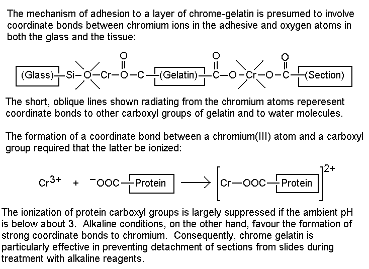
A coordinate bond is a special type of covalent bond fromed between a metal ion and another atom, commonly oxygen or nitrogen. A single chromium(III) ion can form as many a six coordinate bonds. In an aqueous solution of a simple chromic salt the chromium(III) ions are joined by coordinate bonds to the oxygen atoms of water molecules. Strong coordinate bonds can form between chromium and the oxygen atoms of carboxyl groups of proteins. Advantage is taken of this in the process of chrome tanning. Chromium ions cross-link the carboxyl groups of collagen, converting it into leather. Tanning has much in common with histological fixation.
An adhesive for sections is prepared by mixing dilute aqueous solutions of gelatin and chromium potassium sulphate (chrome alum), which is an easily available chromium(III) salt. The resulting liquid has a consistency similar to that of non-drip paint, because the large molecules of gelatin are cross-linked by coordinate bonds to form, perhaps, a single huge molecule. The chrome gelatin cannot be further diluted with water once cross-linking has occurred, which seems to be within a few minutes of mixing the components. Chrome gelatin may be smeared onto glass slides with the end of a finger, or trays of slides may be dipped in the mixture for a few seconds. The treated slides are then allowed to air-dry in a dust-free place. Slides coated with chrome-gelatin are often called "subbed slides." They can be kept for at least three years before using. Sections are floated on water and collected onto subbed slides, which are then allowed to dry in the usual way. Cryostat sections can be collected onto the slides and allowed to thaw and dry. Thicker frozen sections can be mounted from water and adjusted with a paintbrush, but the stickiness of a subbed slide can make it difficult to straighten folds and wrinkles.
The chrome-gelatin solution used for subbing (see BOX 2) quickly becomes infected with micro-organisms. It is therefore made in the laboratory and used the same day.
-------------------------------------------
BOX 2. Making subbed slides.
What you need:
1. Slides (must be clean and dry)
2. Staining racks to hold the slides (glass or metal)
3. Staining tank, just big enough to hold the staining rack
4. Subbing solution. Dissolve 0.3 g gelatin in 290 ml distilled or deionized water. Dissolve 0.03 g chromium potassium sulphate (chrome alum; CrK(SO4)2.12H2O) in 10 ml water (wait; do not heat). Combine the two solutions and pour into the staining tank.
It is advisable to set aside glassware specially for this procedure, because it becomes coated with chrome gelatin, which is difficult to remove.
What to do:
1. Put the clean slides in the staining rack.
2. Immerse in the subbing solution for 30 to 60 seconds, with occasional gentle agitation.
3. Lift out and drain off excess liquid, then leave the rack of slides in a dust-free place until dry.
The dry subbed slides are stored in boxes until they are needed. It is convenient to pack them in the cardboard boxes in which the slides were bought.
Alternative method.
Make up a more concentrated subbing solution (1% gelatin and 0.1% chrome alum.
Smear this over one surface of each slide, and leave the slides to dry, lying on the bench and protected from falling dust.
Pack in boxes when dry.
-------------------------------------------
Protection with a nitrocellulose film.
Nitrocellulose (also called celloidin and collodion, and more correctly named cellulose nitrate) is soluble in ether-alcohol, a mixture of equal volumes of ethanol and diethyl ether. It swells but does not dissolve in ethanol, and is insoluble in 70% alcohol. Undissolved nitrocellulose readily forms a thin membrane, which is permeable to water, inorganic ions and smaller organic ions and molecules such as dyes, but not to macromolecules such as antibodies and other proteins. A properly applied layer of celloidin on top of the mounted sections on a slide can protect against losses even if there is failure of adhesion between the sections and the glass.
The slide bearing the sections must be completely enclosed by the nitrocellulose membrane, with no free edges that might come adrift during processing and staining. In this technique, described in BOX 3, the sections are passed through dehydrating solvents, so the method cannot be used in conjunction with some staining methods, such as those for lipids or most enzymatic activities.
--------------------------------------------------------
BOX 3. Enclosing slides in a nitrocellulose film.
General considerations. This method is applicable to any kind of sections mounted on slides. Paraffin sections must be dewaxed and placed in 100% alcohol (ethanol, methanol or isopropanol). Frozen or cryostat sections must be dehydrated.
What you need:
1. Slides with mounted sections, in 100% alcohol
2. Ether-alcohol.
Diethyl ether (anaesthetic ether is suitable): 500 ml
Ethanol (100%): 500 ml
3. 0.5% Nitrocellulose.
This contains 2.5 g of nitrocellulose in 500 ml of ether-alcohol.
It may be made from a solid form of nitrocellulose (such as parlodion)
or by dilution of a more concentrated solution. Commercial LVN (low
viscosity nitro-cellulose) is sold as a 20% solution.
The 0.5% solution can be kept for a few years, but see safety note below.
4. 70% alcohol. The alcohol concentration is not critical. Add 30 volumes of water to 70 volumes of 100% or 95% ethanol.
What to do:
1. Take the slides to absolute alcohol, in a staining rack.
2. Immerse in the nitrocellulose solution for about 30 seconds, with occasional agitation to ensure that the edges of the slides as well as their surfaces are contacted by the liquid.
3. Lift out the slide rack, shake off excess liquid and wait until they have partly dried by evaporation. This end point is indicated by a change in the lustre of the glass surfaces.
4. Immerse the rack of slides in 70% ethanol for about 2 minutes, to harden the nitrocellulose, then take to water and carry out the staining procedure.
5. Optional. The nitrocellulose film, which is itself stained in some techniques, may be removed. Immerse the slides in ether-alcohol, 2 minutes, after dehydration and before clearing in xylene.
--------------------------------------------------------
Recommended reading.
The following books include detailed discussions of methods for promoting the adhesion of sections to slides.
Culling, C. F. A., Allison, R. T. and Barr, W. T. 1985. Cellular Pathology Technique. 4th ed. Butterworths, London.
Gabe, M. 1976. Histological Techniques (English ed., transl. E. Blackith & A. Kavoor). Masson, Paris.
Kiernan, J. A. 1999. Histological and Histochemical Methods: Theory and Practice. 3rd ed. Butterworth-Heinemann, Oxford.
Lillie, R. D. and Fullmer, H. M. 1976. Histopathologic Technic and Practical Histochemistry. 4th ed. McGraw-Hill, New York.
______________________________________
Updated: September 2004 (to make the graphics readable with Internet Explorer as well as with Netscape).
Updated again: February 2009 (adding a link to the Microscopy Today online archive).