Nanoscale Optical Spectroscopy
Although the past decade has seen rapid developments in the application of nanosized materials such as biological sensors, flexible solar cells, drug delivery carriers, or bit elements in computers, the potential nanoscience holds has yet to be fulfilled. In this context, the characterization of these nano-objects or functionalized surfaces using optical spectroscopy combined with advanced microscopy techniques offers information far beyond that provided by pure imaging techniques as it allows the molecular properties of these materials to be correlated with their molecular structures, sizes and compositions. The possibility to investigate nanomaterials with a spatial resolution much better than 1 micron is a major topic of significance for a better understanding of their optical-mechanical-electrical properties, their interactions and their responsiveness to an external stimuli or perturbation. Similarly, it is important to probe biological processes and chemical exchanges in biological systems. A better spatial resolution or time resolution of the biochemical exchanges would lead to a more precise understanding of the fundamentals of biological processes. Our program focuses on the study of materials and biomaterials organized at the nano- and microscale using a combination of scanning probe microscopy together with a variety of optical microscopy techniques (Raman, Fluorescence). Such an ensemble of complementary methods will be used to (i) determine the confinement effects in semi-conductor nanowires, (ii) develop ultrasensitive vibrational measurements in conjunction with plasmonic platforms and (iii) evaluate chemical exchanges between cells organized on modified surfaces.
Project 1: Tip Raman Enhanced Spectroscopy of Nanomaterials
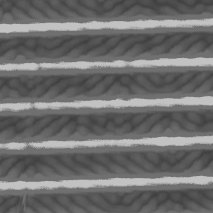
A limitation of optical microscopy is intrinsic to the diffraction limit of light, as stated by the Rayleigh criterion which states that a spatial resolution of lambda/2 can be achieved in the best conditions, lambda being the wavelength of the light. However, this spatial resolution (0.5-1 micron) excludes the precise characterization of individual nanoobjects or domains that are much smaller than the diffraction limit. In this context, tip-enhanced Raman spectroscopy (TERS) employs a metallic tip that scans the near-field of the probed object. The sharp metal tip concentrates the EM field. The optical enhancement is confined at the apex of the metallic tip and allows one to correlate vibrational Raman information with the topography of the object. Using TERS We are investigating a variety of nanomaterials (semiconductors) as well as biomaterials in order to reveal intimate properties at the nanoscale. This work in done in our groups and/or in collaboration with other groups on the material part.
Project 2: Plasmonics Platforms for Optimized Enhancements
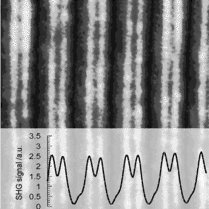
Rapid and accurate identification of trace amounts of chemical species has been a challenge for more than 20 years. The discovery of the surface enhanced Raman effects (SERS) from rough metallic surfaces or colloidal solutions, has stimulated a desire to further increase the detection limit of Raman signals that intrinsically suffer from weak scattering cross sections. Using nanofabrication methods, we have conceived new gold nanostructures which can be effectively used to quantify local enhancements. Such lithographic surfaces composed of noble metals have several advantages. First, their shapes can be well controlled due to the versatility and the reproducibility of nanofabrication tools such as electron beam lithography. My group has recently prepared shapes composed of multiple nanosized triangles using electron-beam lithography and We have shown that the optical properties of such platforms could be finely optimized for maximum enhancement of the electromagnetic field for a determined wavelength. Beyond the fabrication of these platforms we are using them to conduct measurements on a variety of materials using Raman and time resolved fluorescence measurements.
Project 3: In situ Vibrational Spectroscopy of Neurotransmitters in 2D arrays
Background: The spatial control of cell adhesion and growth has made contributions to many different areas including basic cell biology, cell-based biosensors, tissue engineering, design of organ replacements as well as the modeling of cellular interactions. In numerous biological processes such as differentiation and apoptosis, cell-cell interactions such as cell signaling are important and the ability to manipulate these interactions is extremely valuable. Therefore, in order to precisely control the cellular environment, the position of the cells must be manipulated at the singlecell level using surface modification approaches together with microfabrication techniques. When cells are positioned in an ordered arrangement, we can more easily use optical techniques to probe the chemical interactions between adjacent cells using optical microscopy combined with spectroscopy methods. We have developed a new method to prepare such pristine surfaces using plasma deposition of thin films of fluorocarbonpolymers (FC) along with photolithography patterning. The FC film is cytophobic and prevents the adsorption of adhesion proteins, therefore directing cell growth on the more hydrophilic patterned area. Using this method we can organize cells in a defined geometry to study groups and/or individual cells under a confocal microscope using fluorescence or Raman measurements in conjunction with SERS biosensors described in 2. Mamalian cell culture is performed in our cell facility (Director: F. Lagugné) and for more complex cells cultures (neurons) we collaborate with the Robarts Institute.